
The Salicylic Acid Iron III Chloride Test: Why Phenols Turn Violet
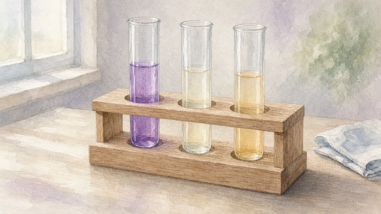
Add a drop of pale yellow iron(III) chloride solution to a little salicylic acid in water and the tube turns violet almost at once. Do the same with clean aspirin and the colour barely changes. That contrast is the whole basis of the salicylic acid iron III chloride test, one of the most familiar quick checks in an undergraduate lab. It’s simple to run, but the chemistry behind the colour is worth a closer look, and so are the limits on what the test can tell you.
What the test looks like
Odinity’s lab reports show the pattern clearly. In one aspirin and salicylic acid lab, the student summarises it this way: “Solutions containing the phenolic group are dark purple and ones without are a yellow color.” Their salicylic acid sample turned purple, while both the crude and purified aspirin stayed yellow-orange. A report comparing stock and student-made aspirin found the same thing. Salicylic acid “dissolved, color turned to purple.” The stock aspirin went orangish-yellow, and the student’s product a light yellow-orange.
That yellow is mostly the iron(III) chloride solution’s own colour. Purdue University’s spectrophotometry handout uses a cuvette of “pale yellow iron(III) chloride solution” as its blank. So a “negative” result isn’t colourless. It’s simply the reagent showing through.
Why phenols and Fe(III) make colour
Salicylic acid carries two groups that matter here: a carboxylic acid and a phenol (an OH attached directly to the benzene ring), sitting next to each other. Iron(III) binds the salicylate through both oxygens. In a 1966 spectrophotometric study in the Bulletin of the Chemical Society of Japan, Ogawa and Tobe wrote the bound ligand as the salicylate dianion, C6H4(O−)(COO−). In other words, the phenol has lost its proton and binds as a phenolate.
The colour comes from that phenolate–iron bond. Many coloured transition-metal complexes owe their colour to faint d–d transitions on the metal. High-spin iron(III) complexes are different: a 2008 study in Chemical Papers of iron(III) complexes of 5-fluorosalicylic acid, a close relative of salicylic acid, notes that those transitions are very weak (molar absorptivity below 1 in the usual units) and are swamped by much stronger bands. In that study’s words, the useful bands are ligand-to-metal charge-transfer (LMCT) bands, “phenolate-O → Fe3+“, lying in the 400–600 nm range. The authors add that their data and the literature on salicylic acid itself show the phenolic OH losing its proton even at very low pH.
In plain terms, visible light pushes electron density from the electron-rich phenolate oxygen toward the electron-poor iron(III). That transition is intense, which is why even small amounts of salicylic acid show up so clearly.
For the 1:1 complex, Ogawa and Tobe found the absorption maximum at 530 nm. That’s green light. A solution that absorbs green looks violet to purple, which is the colour seen in the test tube.
Why aspirin gives so little colour
Aspirin (acetylsalicylic acid) is salicylic acid whose phenol OH has been converted into an acetate ester. How that acetylation happens is covered in Odinity’s post on the salicylic acid and acetic anhydride reaction. For this test, the consequence is what matters: with the phenol capped, there is no phenolate oxygen to bind iron, so there’s no strong charge-transfer band.
Analytical procedures depend on exactly that difference. A University of Central Arkansas lab handout notes that acetylsalicylic acid doesn’t absorb visible light. To measure aspirin by this colour, the handout first boils it with sodium hydroxide, breaking the ester into the salicylate dianion. Only then does the iron(III) reagent turn it violet. Purdue’s procedure follows the same two steps.
The flip side is a useful caution. Anything that breaks aspirin’s ester bond turns it back into a compound that does respond. A faint colour from an aspirin sample means salicylate is present. On its own, that doesn’t say whether the salicylate was left over from the reaction or formed later.
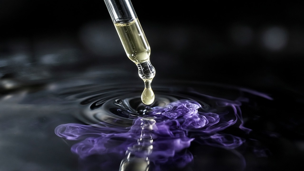
Illustration of violet colour spreading as a drop meets a solution (artist’s impression).
Using the salicylic acid iron III chloride test on aspirin
Qualitative. The everyday version runs side by side. A Louisiana Tech University lab procedure has students add one drop of iron(III) chloride solution to tubes containing water plus a crystal of salicylic acid, a crystal of commercial aspirin, a crystal of their own product, and a reagent-only control. The instruction is explicit: “The presence of unreacted salicylic acid in the synthesized aspirin can be detected with the iron(III) chloride test.” In Odinity’s acetylsalicylic acid lab, the student reports that their product stayed clear, “showing that there were not traces of salicylic acid (which would have turned the compound purple).”
Quantitative. Because the colour’s intensity tracks concentration, the test becomes colorimetry.
- Make a series of standards of known salicylate concentration.
- Add excess iron(III) in acid.
- Read each standard’s absorbance near 530 nm.
- Plot absorbance against concentration to get a Beer’s law calibration line.
- Measure the unknown and read its concentration off the line.
Purdue’s handout uses iron(III) chloride buffered to pH 1.6 with HCl–KCl and reads at 530 nm. In Odinity’s determination of salicylate via reaction with Fe(III), the student built exactly this kind of calibration curve. It was close to linear, with R² = 0.9991. The same student used Job’s method of continuous variation and found maximum absorbance at a 0.5 mole fraction of iron, pointing to a 1:1 iron-to-salicylate complex.
To measure leftover salicylic acid in an aspirin product, rather than total aspirin, the hydrolysis step is skipped. The colour then comes only from salicylic acid that was already free. A medicinal-chemistry practical published by IUPAC describes this route: it determines free salicylic acid in synthesized aspirin from the Fe(III) complex against a standard curve read at 540 nm. That’s slightly off 530 nm. The complex absorbs across a broad band rather than at a single sharp wavelength, so labs differ a little in where they read it. Ultraviolet spectroscopy is another option. In Odinity’s characterization of aspirin lab, the student measured salicylic acid at 298 nm, where aspirin’s molar absorptivity is close to zero.
How pH changes the colour
The test is sensitive to acidity because both the ligand and the iron are. Ogawa and Tobe found the absorption maximum stays at 530 nm between pH 0.7 and 2.5. Above pH 3 it shifts to shorter wavelengths as a second and then a third salicylate binds, giving Fe(Sa)+, Fe(Sa)2− and Fe(Sa)33− in turn. A study in the Journal of Chemical Society of Nigeria located the pH of maximum formation of the 1:1 FeSal+ complex at 2.4–3.0.
Quantitative methods add acid or buffer to the iron reagent. Purdue’s pH 1.6 buffer, for example, sits inside the range where Ogawa and Tobe found a single complex, so the calibration line tracks one species. Odinity’s iron(III) sulfosalicylate stability-constant lab makes the same point for a close relative of salicylic acid. That experiment was run at pH 0–2.5, where the 1:1 complex dominates.
Safety note on the reagent
PubChem’s hazard summary for iron(III) chloride carries the GHS “Corrosive” and “Irritant” pictograms. Of the company reports to the European Chemicals Agency that it aggregates, about 75% classify FeCl3 as causing severe skin burns and eye damage (H314), and about 97% as harmful if swallowed (H302). Even dilute test solutions start from a corrosive solid.
Limits: the test isn’t specific
The violet colour says “a phenol-like oxygen is binding iron.” It doesn’t say “salicylic acid.”
- Other phenols respond too. A qualitative organic analysis procedure on LibreTexts notes that most phenols form coloured complexes with iron(III). The colours vary between red, purple, blue and green depending on the phenol, solvent and concentration.
- A negative result isn’t conclusive. The same LibreTexts procedure warns that “some phenols do not give colours,” so a negative result shouldn’t be taken as significant without supporting evidence.
- Enols respond as well. A George Mason University organic lab handout lists the ferric chloride test for “phenols and compounds with high enolate content.” It attributes the colour to enolate complexation with Fe(III) and notes that the colours can be short-lived.
- Conditions matter. Ogawa and Tobe showed that changing pH changes which complex forms. Two solutions with the same salicylate content can look different if their acidity differs.
In a teaching lab, where the only plausible phenol is salicylic acid, the test is a reliable, fast check for leftover salicylic acid. That is the settled part. The rule of thumb is to treat it as a screen: a calibrated absorbance at a controlled pH tells you how much salicylate is present, and a melting point or spectrum still tells you what the compound actually is.
[…] why a lab checks a product for leftover or regenerated salicylic acid using the colour reaction in the salicylic acid iron(III) chloride test. A similar ester hydrolysis, run deliberately on methyl salicylate, is the first step in […]